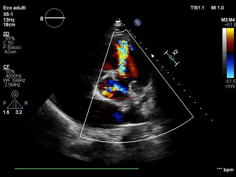
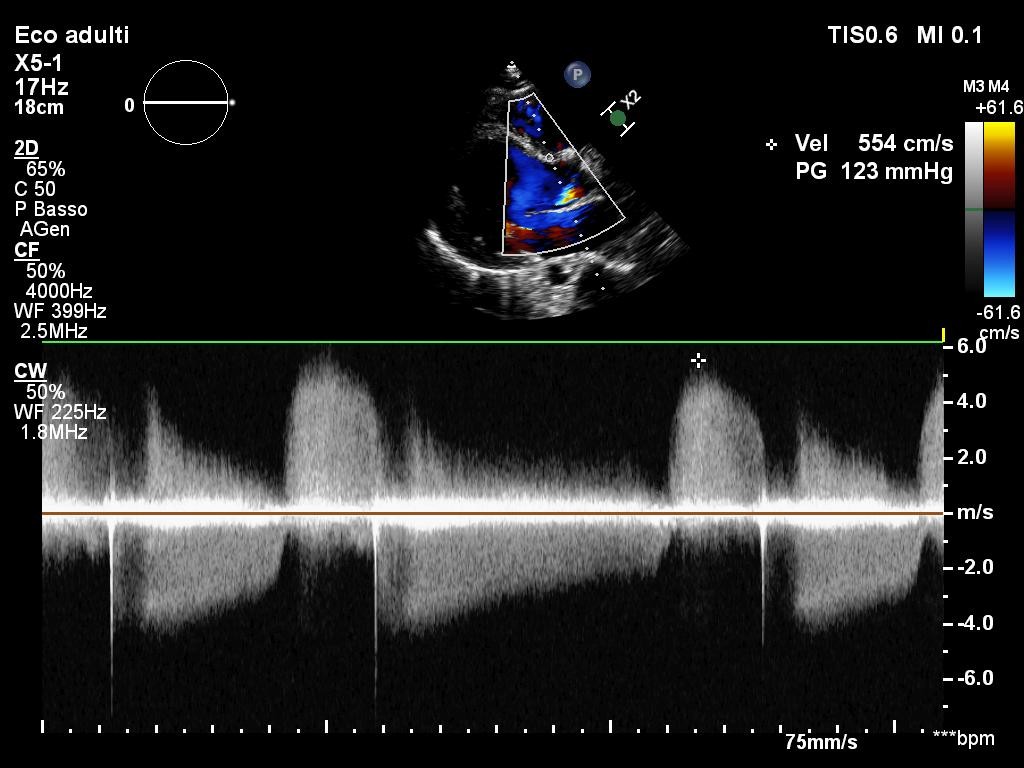
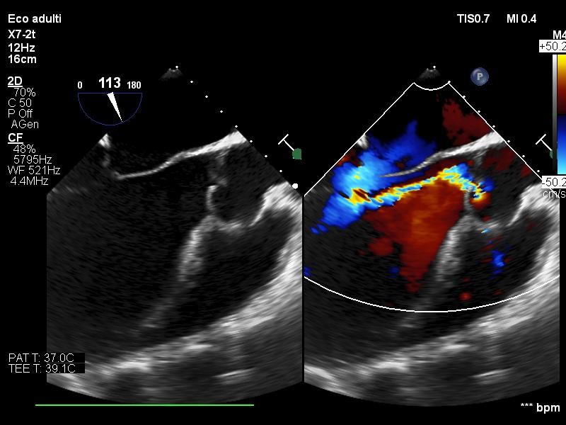
Background. Laubry-Pezzi syndrome is a rare congenital heart disease characterized by the association of a ventricular septal defect (VSD) and aortic regurgitation. Clinical presentation is heterogeneous, and imaging is often crucial for diagnosis . Case description. An 18-year-old Gambian male with no previous cardiac or family history presented to the emergency department with atypical chest pain. The electrocardiogram showed left ventricular hypertrophy, with normal troponin levels and blood pressure. Transthoracic echocardiography (TTE) demonstrated a markedly dilated left ventricle with preserved systolic function, dilation of the aortic root (42 mm), and a restrictive perimembranous VSD with left-to-right shunt (maximum velocity 5.5 m/s, peak gradient 123 mmHg) associated with aortic regurgitation. Transesophageal echocardiography (TEE) was performed for a detailed assessment of the valvular lesion, showing severe eccentric aortic regurgitation with right coronary cusp prolapse, due to the Venturi effect created by the turbulent flow through the VSD. The overall findings supported the diagnosis of Laubry-Pezzi syndrome. The patient underwent surgical repair with pericardial patch closure of the VSD and and mechanical aortic valve replacement. Conclusions. Laubry-Pezzi syndrome, although rare, should be considered in young patients presenting with VSD and aortic regurgitation. Echocardiography remains the cornerstone for diagnosis and surgical planning.