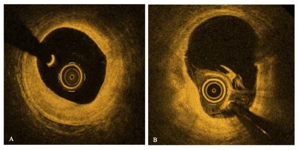
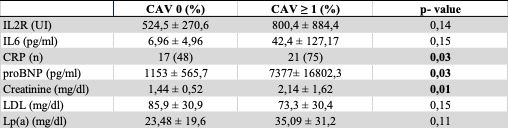
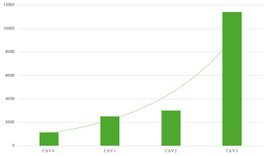
Background: Cardiac allograft vasculopathy(CAV) is the most common form of late rejection after heart transplantation.Early detection and monitoring of CAV are crucial for improving long-term outcomes.Aim of this work is to investigate pros/cons of each imaging modality and the potential of selected non-invasive biomarkers in predicting the presence and severity of CAV. Methods: We retrospectively analyzed clinical reports and angiographies of 63 living patients transplanted in Ospedale Monaldi. Levels of NT-proBNP, creatinine, LDL,Lp(a),IL-2R, IL-6, C-Reactive protein were statistically analyzed. Results: sample was stratified as follows:56% CAV0, 9% CAV1, 11% CAV2 and 24% CAV3 disease. IVUS(n=29)resulted positive in 67% cases of CAV0group, identifying a pre-clinical stage of disease.OCT(n=2)resulted positive in both patients, showing in one case a fibrotic infiltration of the vessel wall, diffusely involving the entire analyzed segment(Figure1A), and in the second case lipid accumulation at multiple sites along the vessel(Figure1B). Results about biomarkers are summarized in Figure2.CRP,NT-proBNP, and serum creatinine were significantly correlated with angiographically confirmed CAV(Figure3),whereas IL-2R and IL-6 showed a trend toward higher values in the CAV group. No statistically significant difference was found in mean LDLand Lp(a) levels between CAV0 and CAV-positive patients.No difference was found for all the aforementioned parameters between IVUS-pos and IVUS-neg patients. Discussion :the main findings of our study are the following:1)Intravascular imaging currently remains the sole system for preclinical diagnosis of the disease;while IVUS discriminates the presence of disease, OCT can reveal different disease phenotypes;2)CRP showed a statistically significant correlation with disease presence,while IL-2R and IL-6 exhibited an increasing trend with disease severity, confirming the role of inflammation in the development of CAV;3)no statistically significant differences were found for LDL or Lp(a) levels between groups, which underscores that CAV is not purely an atherosclerotic process;4)higher creatinine and NT-proBNP levels may reflect allograft dysfunction or ongoing vascular pathology in CAV patients. Conclusions :This study highlights the potential clinical value of inflammation, renal dysfunction and myocardial stress biomarkers in CAV patients, and advocate for an expanded use of intravascular imaging, especially OCT, for early CAV detection.