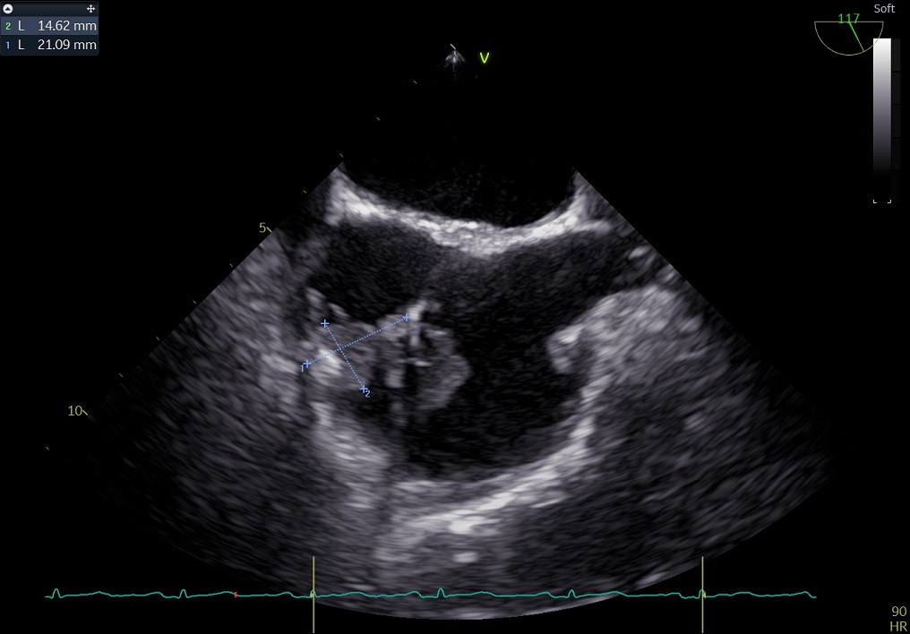
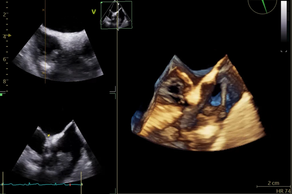
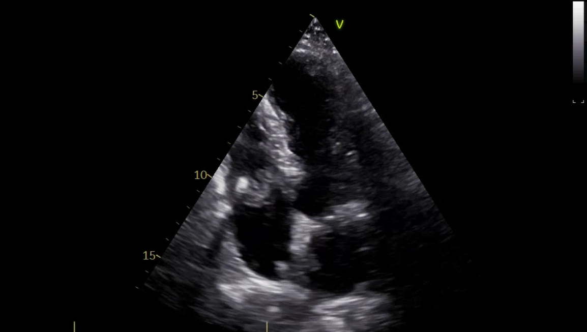
Cardiac implantable electronic device (CIED)-related infective endocarditis represents a diagnostic and therapeutic challenge, particularly in the presence of negative blood cultures. We report the case of a 60-year-old woman with a history of resuscitated cardiac arrest, idiopathic myelofibrosis, hypothyroidism, and implantable cardioverter-defibrillator (ICD) implantation, who was admitted for persistent fever despite empirical antibiotic therapy. Physical examination revealed signs of device pocket infection, while laboratory tests showed marked systemic inflammation; repeated blood cultures were negative. Transthoracic and transesophageal echocardiography demonstrated a large (28×11 mm), highly mobile vegetation with high embolic potential attached to the ventricular lead of the ICD, without significant valvular involvement. Given the strong clinical suspicion, 18F-FDG PET-CT was performed and revealed pathological tracer uptake at the device pocket, allowing a definitive diagnosis of CIED-related endocarditis according to the 2023 ESC criteria, despite persistently negative blood cultures, likely influenced by prior antibiotic exposure. The patient was treated with empirical antibiotic therapy and underwent complete device extraction using a hybrid angio-VAC–assisted procedure, with a favorable outcome. After normalization of inflammatory markers and persistently negative blood cultures, a subcutaneous ICD was implanted. The in-hospital course was complicated by acute pulmonary embolism in the setting of SARS-CoV-2 infection, confirmed by CT pulmonary angiography and treated with oral anticoagulation and antiviral therapy. A repeat PET-CT scan excluded an infectious nature of the pulmonary thrombus. After a total of six weeks of antibiotic therapy, the patient was discharged in good clinical condition. This case highlights the pivotal role of multimodal imaging in the diagnostic workup of CIED-related endocarditis, the importance of early and complete device removal, and the need to consider SARS-CoV-2 infection as a prothrombotic risk factor.