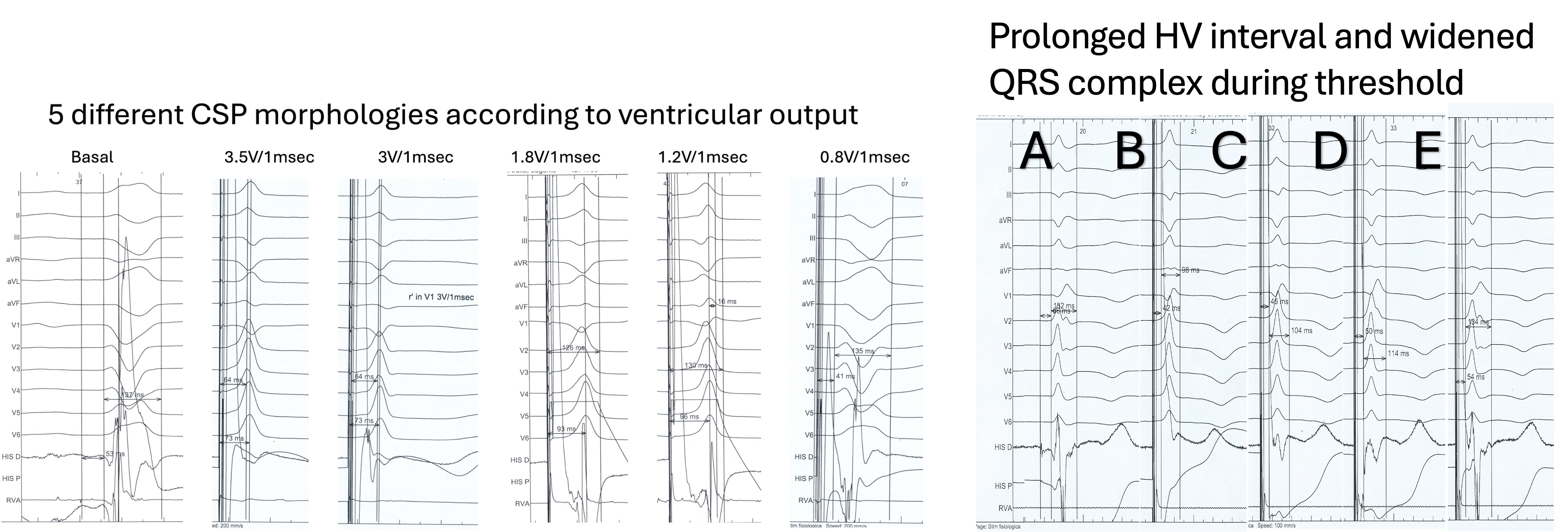
Background: His bundle pacing (HBP) is considered the most physiological method for cardiac pacing. In heart failure patients with narrow QRS, we noted varying clinical effects depending on HBP output (HIPPOS study ongoing). Here, we report two cases demonstrating how different conduction velocities correspond to different intensities of HBP output. Methods/Results: An 82-year-old woman presented with high-grade AV block and narrow QRS. During lead fixation showed initial RBBB morphology, which transitioned to LBBB with stable AV conduction. High output HBP (3.5V/1ms) corrected LBBB . At 3V/1ms an r’ in V1 was observed with a small increase in HV from 44 to 46ms, while at 1.8V/1msec there was a “jump” from 74 to 92ms in RWPT in V6, loosing however the r’in V1. QRS was larger from 74 to 88msec. A s output decreased, QRS widened and HV interval varied, with selective HBP at low outputs failing to correct LBBB. In a 76-year-old man with AF and spontaneous RBBB, HBP reduced QRS duration and HV interval as output increased. Lower voltages led to longer QRS and HV intervals, again resulting in selective HBP without RBBB correction before threshold. Results: In both cases, the variations in pacing intensity revealed distinct changes in ECG morphology and conduction intervals, highlighting the dynamic relationship between HBP output and myocardial conduction. These observations suggest that incremental adjustments in pacing voltage can modulate the degree of bundle branch block correction, as reflected by alterations in QRS duration, HV interval, and waveform characteristics. Conclusions: In both cases there is evidence of conduction system capture from the highest to the lowest voltage. Pacing from high to low voltage in both cases produced multiple ECG morphologies, with evidence that higher HBP outputs result in greater conduction velocity and more complete bundle branch block correction.