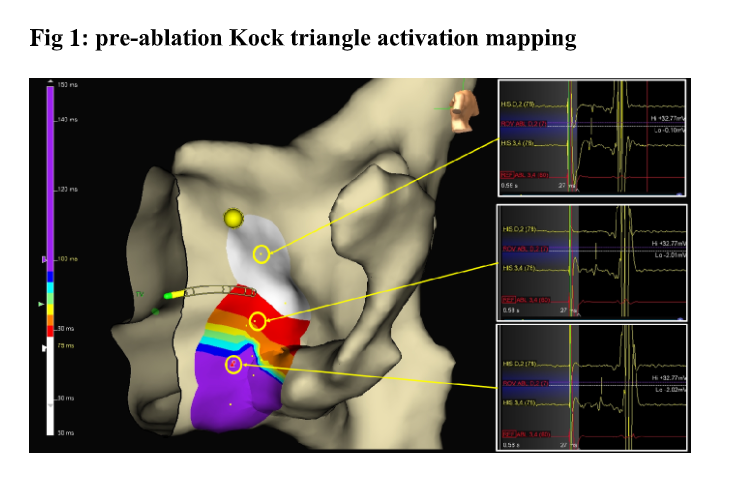
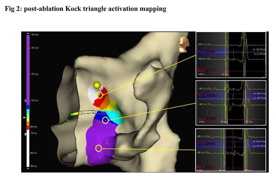
Introduction Slow pathway ablation is currently the target for AV nodal reentrant tachycardia (AVNRT) with a high success rate of almost 100% and 0.5% to 2% risk of II- to III-degree AV block. Kock triangle (KT) mapping is helpful to define the fast pathway location, to find out its physiologic anteroseptal location. We aimed to describe the KT mapping by means of electroanatomic (EA) activation mapping of fast pathway, performed beside the conventional EP study. Methods KT was mapped as follows: atrial pacing from peri-Hisian antero-septal (AS), mid-septal (MS) and postero-septal (PS) regions, by means of 4 mm ablating catheter beside a quadripolar diagnostic catheter placed on His region. The EA local activation time (LAT) was performed by means of Ensite X system (Abbott Medical) in all patients; the window of interest was set from 10 msec after the pacing atrial spike extended beyond the ventricular EGM on the His catheter tracing. The spike- to-His deflection (S-H) interval was measured to define the fast pathway location (activation gradient). On the ablator catheter tracing the annotation was set (ROV), timed to the His deflection on His catheter tracing; the annotation was set even when no His deflection was recorded on ablator tracing as in MS and PS regions (“mirroring”). The KT was mapped before and after successful elimination of the slow pathway. Results Seventeen consecutive AVNRT patients were included who underwent to successful ablation with slow pathway elimination. In all cases, at baseline pre-ablation KT mapping, a progressive prolonged S-H interval was found (AS<MS<PS), depicted as activation gradient, traditionally meaning as anteroseptal fast pathway physiologic location and confirmed by the EA LAT (Fig 1). As compared to pre-ablation, the post-ablation LAT mapping showed the same activation gradient in all patients (Fig 2), as follows: 1) pre-AS= 89,2 msec vs post-AS= 91,2 msec p=0.2 2) pre-MS= 106,8 msec vs post-MS= 110,2 msec p=0.15 3) pre-PS= 127,7 msec vs post PS=130 msec p=0.38 (Normality of paired differences was assessed using the Shapiro-Wilk test and was not violated; therefore Student’s t- test was applied) Conclusion The activation mapping of KT by LAT may help to define the AV node dual physiology beside the conventional EP mapping. It might depict a safer region for slow pathway ablation and confirms the preserved anterograde conduction properties of fast pathway after slow pathway ablation.