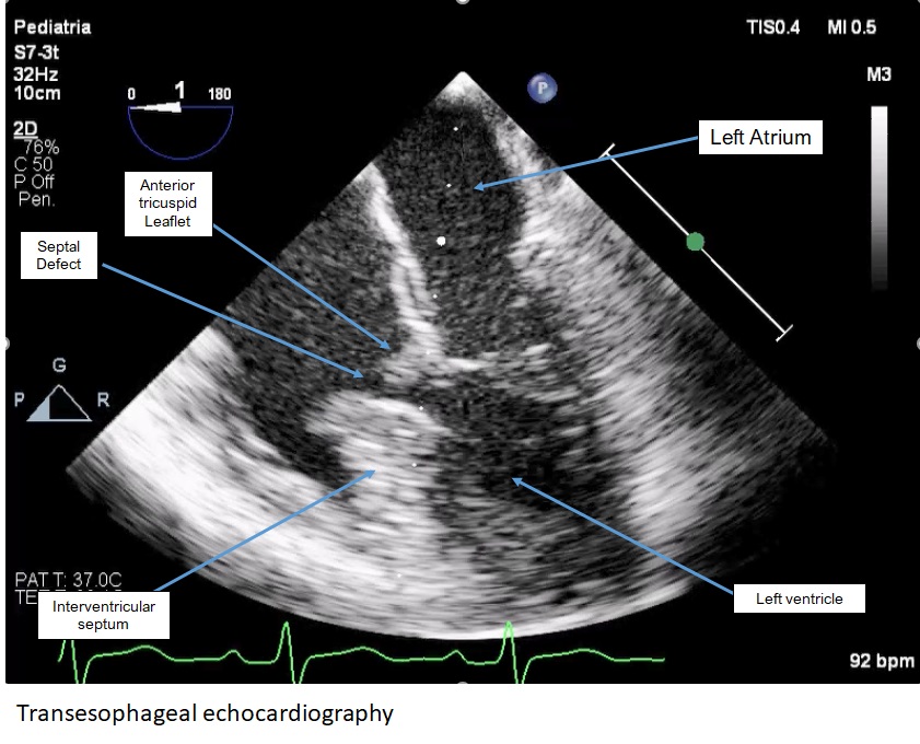
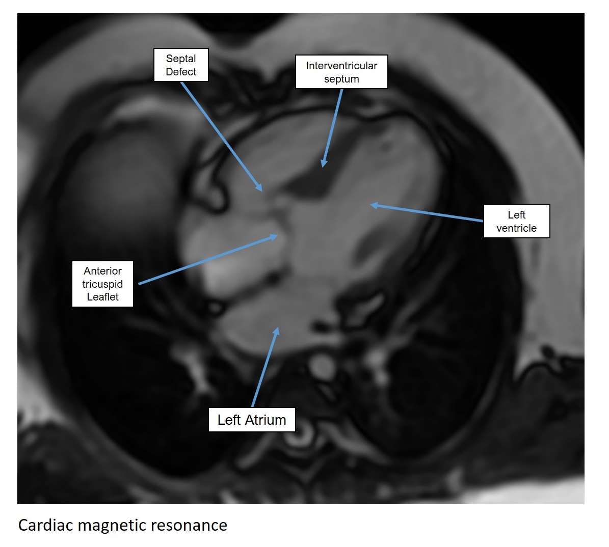
Introduction Tricuspid valve overriding is a rare congenital anomaly of the atrioventricular junction, frequently associated with a ventricular septal defect. Differentiation from more complex conditions, such as tricuspid straddling or unbalanced atrioventricular septal defects, is crucial for accurate prognostic assessment and to avoid unnecessary therapeutic interventions. In this setting, multimodality imaging plays a central role. Case report We report the case of an 8-year-old boy referred to our center after the incidental detection of a cardiac murmur following Parvovirus B19 infection. Electrocardiography revealed sinus rhythm with incomplete right bundle branch block. Transthoracic echocardiography showed posterior interventricular septal malalignment with tricuspid valve overriding and a perimembranous ventricular septal defect partially closed by the tricuspid subvalvular apparatus, associated with a mild left-to-right shunt (Qp/Qs ≈1.4) and mild tricuspid regurgitation. Biventricular systolic function was preserved. Holter monitoring and exercise testing showed no arrhythmias and good age-related functional capacity. Cardiac magnetic resonance imaging confirmed tricuspid valve overriding with a small perimembranous ventricular septal defect (≈5 mm), a very mild interventricular shunt (Qp/Qs = 1.2), normal biventricular volumes and systolic function, and no evidence of intramyocardial fibrosis. Transesophageal echocardiography definitively excluded tricuspid valve straddling, allowing complete anatomical characterization. Discussion Anomalies of the right atrioventricular complex encompass a wide morphological and functional spectrum. In patients with mild shunting and preserved ventricular function, distinguishing simple tricuspid overriding from more complex forms is essential. The integration of transthoracic and transesophageal echocardiography with cardiac magnetic resonance enables accurate anatomical definition, reliable shunt quantification, and appropriate prognostic stratification. Conclusions This case highlights the key role of multimodality imaging in the management of rare congenital heart diseases involving the atrioventricular junction. Precise anatomical assessment supports a conservative management strategy and tailored follow-up in asymptomatic patients with mild shunt and normal ventricular function, avoiding unnecessary invasive interventions.