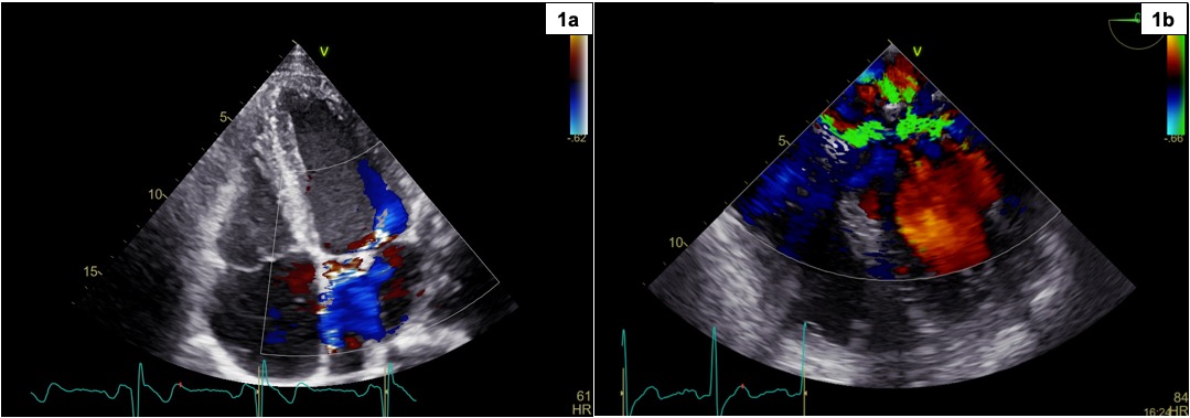
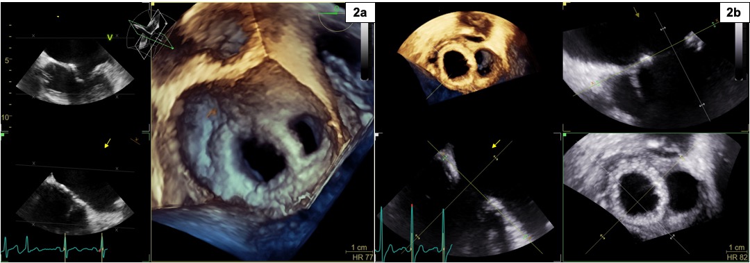
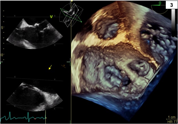
INTRODUCTION: Annuloplasty is one of the main mitral valve repair techniques for the treatment of severe insufficiency. One of its potential complications is the detachment of the prosthetic ring, resulting in recurrence or worsening of regurgitation. CLINICAL CASE: A male patient with Marfan syndrome underwent at the age of 15 to mitral annuloplasty for severe regurgitation associated with Barlow's disease and to valve sparing ascending aorta replacement (David technique) for root aneurysm and severe valvular regurgitation. At the age of 20, TTE and 2D-TEE revealed detachment of the medial portion of the mitral prosthetic ring, with recurrent moderate regurgitation overall (1a) and evidence of a jet that divided immediately after its origin into a medial component, stretched between the native annulus and the prosthetic ring, and a more central component (1b). The regurgitation was contained by the non-coapting portion of the prosthesis, which limited the systolic motion of the redundant valve leaflets, preventing higher-grade regurgitation. An endocarditis as cause of the detachment was ruled out, which was attributed instead to the connective tissue laxity of Marfan patients. In agreement with the cardiac surgeons, close follow-up was started. With the advent of 3D technology, follow-up TEE confirmed the presence of a wide detachment of the medial portion of the prosthetic ring, which arched over the native valve and caused a double-orifice appearance during diastole (2a, 2b) and limited the movement of prolapsing leaflets during systole, restricting the degree of regurgitation (3). In subsequent years, the mitral regurgitation remained moderate, while the aortic regurgitation worsened to severe. At the age of 30, the patient developed dyspnea associated with initial dilation and reduced LVEF, prompting mitral-aortic valve replacement with mechanical prostheses. The follow-up TTE showed normalization of both LV dimensions and EF. DISCUSSION: Mitral prosthetic ring detachment with recurrent regurgitation is a rare but possible complication that does not always require immediate intervention. 3D-TEE is now a valuable tool as it provides an image of the entire valvular area, allowing for better anatomical-functional characterization and aiding in differential diagnosis with other conditions, such as infective endocarditis. CONCLUSION: 2D and 3D echocardiographic imaging enabled an accurate diagnosis and guided the appropriate therapeutic approach.