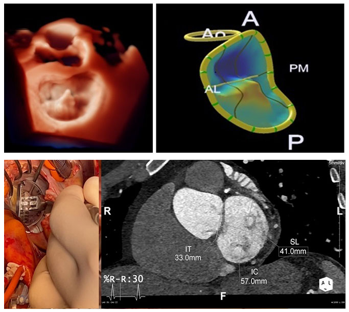
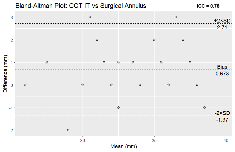
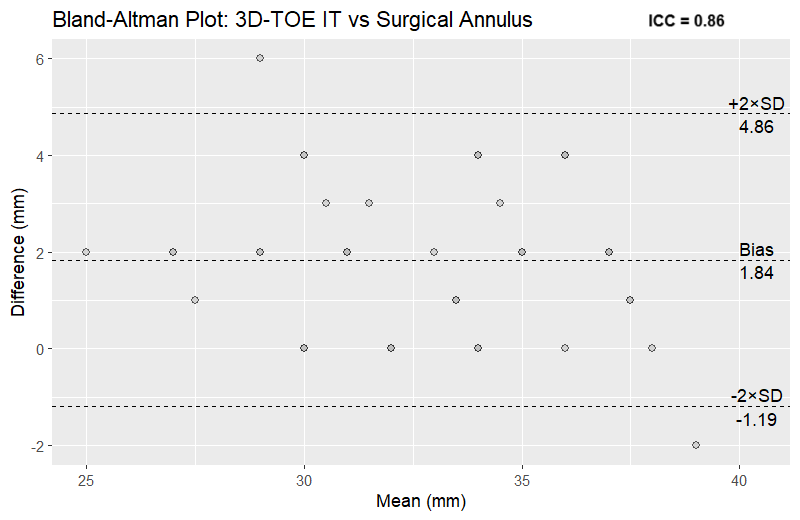
BACKGROUND: The success of mitral valve repair with annuloplasty depends on the repair technique and the correct sizing of the mitral annulus ring. Three-dimensional transoesophageal echocardiography (3D-TOE) has been proven to be essential for the anatomo-functional characterization of mitral valve apparatus in patients undergoing surgically mitral valve repair. It also allows the measurement of quantitative parameters useful in determining the size of the annuloplasty ring. Cardiac computed tomography (CCT) plays a key role for device sizing in patients undergoing transcatheter mitral valve replacement; it provides isotropic sub-millimetre spatial resolution and it is the gold standard for geometric characterisation of the mitral valve and for assessment of its spatial relationship to adjacent anatomical structures. PURPOSE: To compare intra-operatively 3D-TOE assessment of mitral valve apparatus with CCT images and to compare 3D-TOE and CCT measures with intraoperative surgeon-measured mitral valve annulus. METHODS AND RESULTS: We used a cohort of 39 patients who underwent surgery for severe primary mitral regurgitation with mitral valve repair with annuloplasty, all of whom had undergone CCT prior to surgery. We compared measurements of the mitral annulus (intertrigonal distance and intercommissural distance) obtained by 3D-reconstruction of intra-operative TOE and CCT images with the ring measured intraoperatively by the surgeon and then implanted, using intraclass correlation and Bland-Altman analysis. We found that intertrigonal distance measured by both CCT (ICC 0.78 [CI 0.62-0.88; p < 0.001]) and 3D-TOE (ICC 0.86 [CI 0.75-0.92; p < 0.001]) showed a good reliability with the surgical ring; only intercommissural distance measured by 3D-TOE showed a good reliability with the surgical ring (ICC 0.78 [CI 0.62-0.88; p < 0.001]), while the same measure obtained by CCT showed a moderate reliability (ICC 0.60 [CI 0.32-0.78; p < 0.001]). We then compared the intraoperative 3D TOE measurements with those obtained by CCT and found excellent reliability when comparing the intertrigonal distance (ICC 0.94 [CI 0.88-0.97; p<0.001]). CONCLUSIONS: 3D-TOE can be considered as a first-line imaging technique to assess the anatomical features of the mitral valve apparatus, not only in patients undergoing cardiac surgery, but also in those undergoing percutaneous interventions or with contraindications to contrast medium CT.