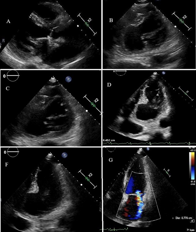
The image shows a giant pseudoaneurysm of the basal and mid segments of inferior portion of the septum and of the inferior wall of the left ventricle as a late complication (after about one mount) of a subacute myocardial infarction in 70-year-old woman, underwent percutaneous revascularization of a totally occluded right coronary artery [1]. The patients underwent left ventricular plastic surgery and mitral valve replacement with a biological prosthesis. Figure legend: transthoracic echocardiographic images showing in (A) parasternal long axis view on left ventricle, left atrium e the first portion of the ascending aorta. Panels B e C shows in parasternal short axis views the giant pseudoaneurysm involving the inferior portion of the septum and of the inferior wall of the left ventricle and the posteromedial portion of the mitral valve apparatus. It is possible to observe the narrow neck (white arrow), the ventricular free wall rupture contained by localized pericardial adhesions (double white arrow). Pannels D and F show an apical 4 chamber view with the pseudoaneurysm of the mid and basal portion of inferior septum. Severe mitral regurgitation due to the disruption of mitral valve apparatus was also present, as shown in panel G.