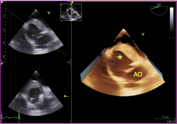
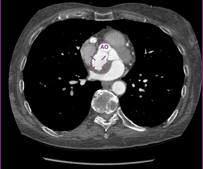
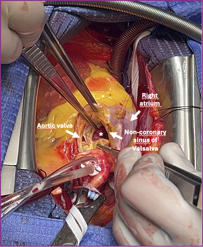
Clinical Presentation A 68-year-old woman was referred to our attention due to dyspnea on exertion. She denied cardiovascular risk factors, chest pain, syncope, and palpitations. 2D-transthoracic echocardiography showed normal systolic and diastolic function of the left ventricle with mild aortic regurgitation and a partially vacuolated mass (31×33 mm) attached to the interatrial septum. Clinical Investigations The hypothesis of atrial myxoma was assessed by transesophageal echocardiography, revealing an echo-dense and partially liquified structure at the level of the non-coronary sinus of Valsalva, compressing and deviating the interatrial septum; mild aortic regurgitation was confirmed. 3D analysis showed an aneurysmal sac originating from the non-coronary sinus of Valsalva, suggesting the hypothesis of a pseudoaneurysm. Negative inflammatory markers and blood cultures ruled out the aneurysmal evolution of a septic abscess. Thoracic CT showed a large pseudoaneurysm (34x28mm) of the non-coronary sinus with eccentric thrombotic stratification, impinging on the interatrial septum. Management The clinical case was evaluated by the Heart Team, and the decision to perform surgery for pseudoaneurysm exclusion was made. The surgical view showed a large defect in the aortic wall at the non-coronary sinus of Valsalva, resulting in a huge pseudoaneurysm with extensive thrombus. Exclusion of the pseudoaneurysm was performed, followed by reconstruction of the aortic wall using a heterologous pericardial patch. The postoperative course was uncomplicated. Since no secondary causes have been observed, genetic evaluation has been indicated, but results are still ongoing. Discussion Aneurysms of the sinus of Valsalva (SOVAs) are rare, with an estimated prevalence of 0.09% in the general population. The right coronary sinus is the most frequently involved, followed by non-coronary and left coronary sinus. Endocarditis and acute aortic syndromes are responsible for acquired SOVAs. Congenital aneurysms result from an incomplete fusion of the aortopulmonary septum with the interventricular septum and are often associated with Marfan syndrome or other genetically determined connective tissue disorders. Conclusions This clinical case indicates that spontaneous pseudoaneurysms are a rare but not impossible finding. After ruling out infectious or traumatic causes, a genetic origin should be considered even in patients without distinct features of collagen disease.