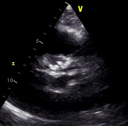
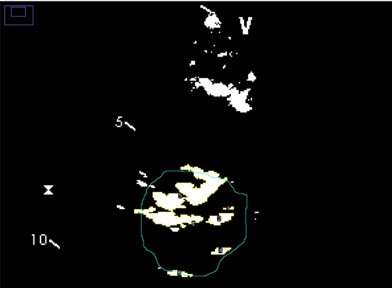
Transthoracic Echocardiography is the main imaging technique for severity grading of Aortic Valve stenosis by measuring peak transvalvular flow velocity, peak and mean transvalvular pressure gradient and Aortic valve area, usually through continuity equation. There is often discordance between Aortic valve area and transvalvular peak velocity and pressure gradient, requiring CT derived aortic valve calcium score in order to discriminate true severe Aortic stenosis from pseudosevere, especially in the case of low-flow low-gradient paradoxical severe Aortic stenosis. Previuosly aortic valve calcium measure on TTE image was attempted by 2D image binarization and a grey scale pixel value of more than 130 has been experimentally used to indicate calcium (Gillis K et al PLOS ONE 2015) Methods: We enrolled 20 consecutive patients (10 men) admitted to our Hospital from 1th September 2025 to 30th of November 2025 with Aortic valve stenosis (Vmax > 2.6 m/sec). Transvalvular Doppler parameters and Aortic Valve Area were aquired for each patient, moreover on a 2D image frame from parasternal short axis view of the aortic valve (Figure 1) image binarization was performed (Figure 2) using a free online software for image elaboration (ImageJ, https://imagej.net/ij/) and calcium threshold for binarization was determined as 130 + m, where m was mean grey scale value from RVOT "black" blood pool, finally total calcium area of the aortic valve was manually tracked and measured in square cm using the above mentioned software and the so obtained value x 1000 was denominated EAU (echo agatston units), so that 1 cmq corresponded to 1000 EAU. Results: mean +/- sd were: EF (59.9 +/- 5.9%), AVA (0.67 + 0.27 cmq), Vmax (4.146 + 0.86 m/sec), mean gradient (45.77 +/- 21.22 mmHg), dimensionless index (0.23 +/- 0.09), EAU calcium score ( 1240 +/- 792 EAU). A significant negative correlation by Pearson correlation score was found between EAU Calcium Score and Aortic valve area by continuity equation (r: -0.574 p <0.01), there was also a significant negative correlation between EAU Calcium score and DVI (r: -0.578 p <0.01) and a weak but significant positive correlation between Echo Calcium score and peak velocity (r: 0.483, p: 0.03) and mean gradient (r: 0.470 p: 0.036) Conclusion: Aortic Valve Calcium Score measure from 2D Echocardiography is feasibile with a free online software and can be used as an adjunctive parameter for severity grading of aortic valve stenosis