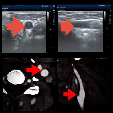
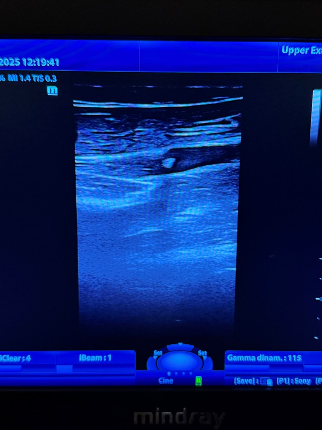
Carotid web is a rare abnormality of the arterial wall, considered a variant of intimal fibromuscular dysplasia, and has been associated with cryptogenic ischemic stroke in young adults. Its detection by Doppler ultrasound of the supra-aortic trunks remains a diagnostic challenge, as it is often under-recognized and misinterpreted as artifacts or thin atherosclerotic plaques. We report the case of a 35-year-old woman, asymptomatic at vascular follow-up, in whom ultrasound revealed a thin intraluminal membrane at the carotid bulb, with the characteristic "shelf-like" appearance and a "fish-mouth" pattern. This case raises questions about the management of asymptomatic patients and the indications for second-level imaging or preventive treatment. History: A 35-year-old woman, with no cardiovascular risk factors, under routine vascular follow-up for a family history of atherosclerosis. No history of cerebrovascular events or neurological symptoms. Physical examination: Normal blood pressure, no vascular bruits. Neurological examination within normal limits. Ultrasound findings: – B-mode imaging of the supra-aortic trunks revealed a thin, hyperechoic intraluminal membrane protruding from the posterior wall of the ICA bulb, without crossing the lumen entirely. – Transverse view showed the classic "fish-mouth" appearance. – Color Doppler demonstrated laminar flow in the common carotid artery, but local turbulence and small areas of vortex stagnation adjacent to the membrane. – No hemodynamically significant stenosis was detected (ICA PSV < 120 cm/s). Initial follow-up: The patient remained asymptomatic. A CT angiography was recommended for diagnostic confirmation and risk stratification. This case highlights that carotid web may be incidentally identified in young asymptomatic patients. Accurate diagnosis is essential to differentiate it from ultrasound artifacts or thin atherosclerotic plaques. In asymptomatic individuals, management remains debated: currently, the most widely accepted approach involves clinical and ultrasound surveillance combined with antiplatelet prophylaxis, with second-level imaging indicated for confirmation. Prospective studies are needed to define risk criteria and optimal therapeutic strategies.