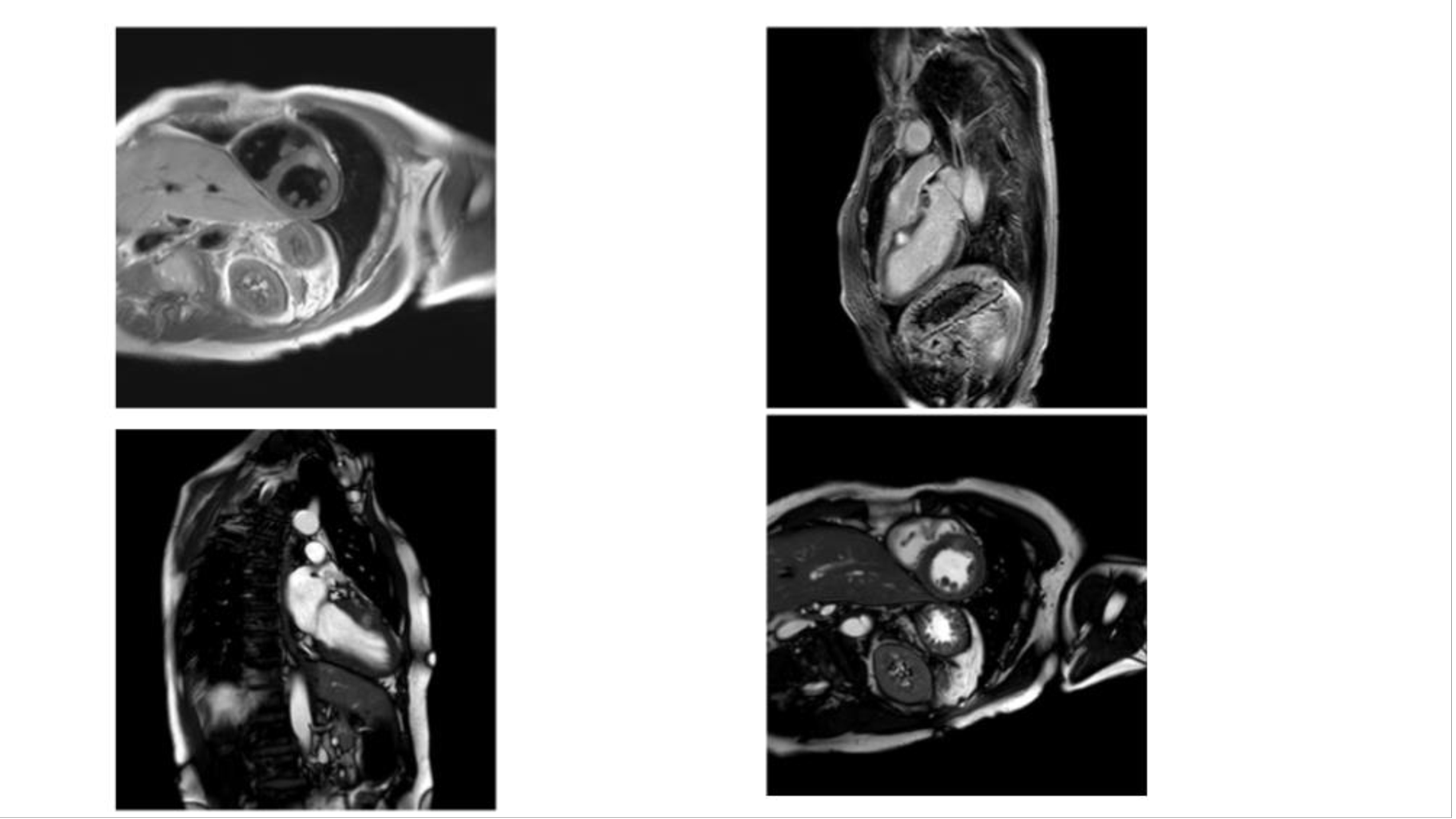
Patient presentation: we present the case of a patient with a recent melanoma metastasis to the left ventricle (LV). The patient is a 57-year-old male who, in March 2013, underwent excision of a pigmented lesion of the right hip with histological diagnosis of superficial spreading melanoma. In July 2024, a follow up computerized tomography (CT) scan showed nodular lesion suspect for metastasis. Patient thus underwent splenectomy with pathological confirmation of melanoma metastasis, BRAF V600E mutation. Adjuvant Nivolumab was administered. In February, the patient routinely performed a CT scan that showed melanoma recurrence with lymph nodes and lung involvement. An urgent positron emission tomography (PET) with 18-fluorodeoxyglucose (FDG) scan was performed and revealed multifocal, intensely avid, metastatic deposits. In addition, focal uptake was seen in the LV myocardium. Diagnosis and management: the patient was sent for cardiac magnetic resonance (CMR) that revealed well-defined iso-to-hypointense (melanin-containing) homogenous masses in LV (13 x 16 millimeters). It showed increased values on T1 and T2 mapping, as well as increased signal intensity on T2-weighted imaging and enhancement on late gadolinium enhancement images. The findings are highly suggestive of melanoma metastasis. Taking into consideration the primary resistance to anti-PD-1 moloclonal antibodies, patient was promptly commenced on targeted therapy with BRAF tyrosine kinase inhibitor, Encorafenib and MEK inhibitor, Binimetinib. After two months of treatment, the patient showed a reduction in 18-FDG uptake in the myocardium and at other sites of secondary lesions on the 18-FDG PET scan. Conclusions: cardiac metastases from melanoma, before the advent of immunotherapy, were treated with palliative therapy. However, with the advent of immunotherapy, some cases are treated with immunotherapy with excellent results. Patients with BRAF-mutated melanoma tend to respond very quickly to treatment with RAF and MEK inhibitors. Given the primary resistance to anti-PD-1 monoclonal antibodies and the life-threatening nature of the LV metastasis, the patient was promptly started on targeted therapy with BRAF tyrosine kinase inhibitor, Encorafenib, and MEK inhibitor, Binimetinib. This case shows a great response to targeted treatment and proves how medical treatment without surgery can drastically reduce a malignant tumor's mass