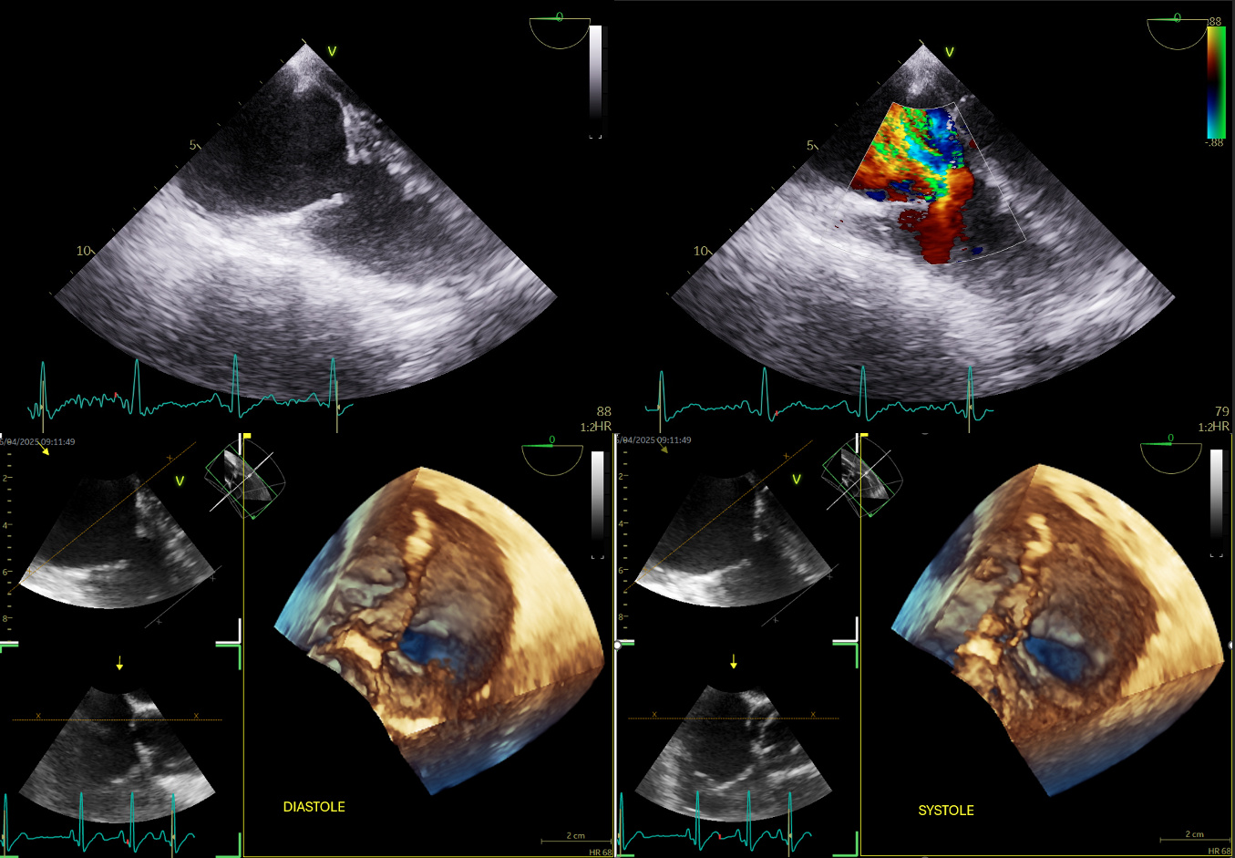
BACKGROUNG Significant tricuspid regurgitation affects 3–6% of the general population and is associated with reduced quality of life, increased hospitalizations and mortality. Echocardiography is the first-line imaging modality for assessing etiology, mechanism, and severity. CLINICAL CASE We describe a 58-year-old woman with hypertension and active smoking, without significant prior cardiac history, who presented with progressive lower-extremity edema and exertional dyspnea. Transthoracic and transesophageal echocardiography showed a normal left ventricle with preserved systolic function and a diastolic D-shaped septum. The tricuspid valve exhibited leaflet fibrosis, retraction, and reduced mobility (area 3D 2,7 cmq), resulting in severe regurgitation (EROA 3D 2,3 cmq) with right atrial and caval dilation. Right-heart catheterization confirmed severe tricuspid regurgitation with markedly elevated central venous pressure, normal pulmonary pressures, and no increase in pulmonary vascular resistance. Contrast-enhanced CT revealed a hepatic mass, diagnosed as a neuroendocrine tumor on biopsy. The characteristic valvular abnormalities raised suspicion for carcinoid heart disease. Ga-68 PET/CT demonstrated pathological tracer uptake in the hepatic lesion, while the primary tumor site remains unidentified. Therapy with a somatostatin analogue was initiated after multidisciplinary evaluation. The patient recently underwent tricuspid and pulonomary valve replacement with a biological prosthesis (respectively Magna 25 and Inspiris resilia 19); the procedure was complicated by advanced atrioventricular block, which required implantation of a Medtronic Micra DR leadless PMK. During the follow-up, although brief, the patient maintained good hemodynamic control with clinical and laboratory improvement. The echocardiogram showed a well-functioning tricuspid and pulmonary bioprosthesis with normal antegrade gradients (max 10 mmHg and mean 4 mmHg) with no valvular regurgitation. DISCUSSION Carcinoid tumors are uncommon neuroendocrine neoplasms producing vasoactive substances that induce fibrotic plaque deposition on right-sided cardiac valves, leading to regurgitation or stenosis. Patients typically present with dyspnea, fatigue, and signs of right-sided volume overload. CONCLUSIONS Tricuspid regurgitation may arise from various mechanisms; carcinoid heart disease represents a rare (but pathognomonic) etiology with distinctive echocardiographic hallmarks.