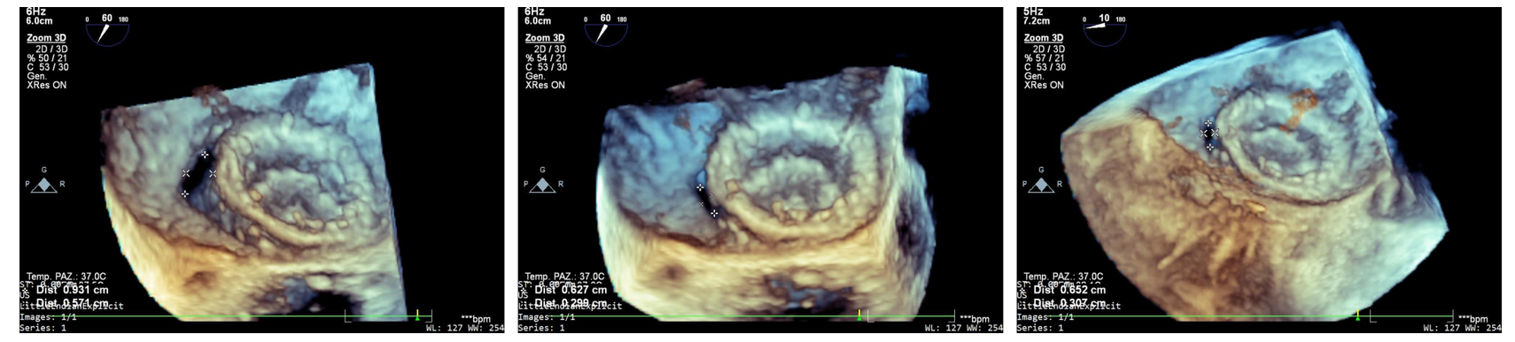
Background Paravalvular leak (PVL) is a complication of valve replacement with 1% to 3% of patients developing symptoms including heart failure or haemolysis. The correlation between the size of the leak and the severity of haemolysis is unclear. Small leaks can cause severe haemolysis, whereas significant leaks may cause no haemolysis. Methods We report the case of an 85-year-old woman who underwent a mitral valve replacement thirty years ago with a mechanical Sorin 27 valve prosthesis. The patient presented at our hospital with chest pain, dyspnoea, cutaneous jaundice and dark-coloured urine. Laboratory tests revealed severe haemolytic anaemia (haemoglobin 76 g/L), markedly elevated LDH (3789 U/L), indirect hyperbilirubinaemia (4.76 mg/dL) and undetectable haptoglobin; transoesophageal echocardiography described a mild-to-moderate anterolateral paravalvular leak (vena contracta 0.2 cm²) at 11 o’clock position, with normal functioning of the bioprosthesis. Extensive diagnostic workup was performed to exclude alternative aetiologies, including abdominal CT and abdominal ultrasound, cytofluorimetry (to exclude paroxysmal nocturnal haemoglobinuria) and haematologic consultation to exclude autoimmune and infectious diseases. Leak correction was performed through percutaneous implantation of two Amplatzer Valvular Plug III devices, resulting in clinical improvement and haemodynamic stabilization. Results The postoperative course was characterized by marked improvement in symptoms, physical findings (resolution of jaundice and urine colour), and laboratory parameters. Haemoglobin stabilized around 90–92 g/L, LDH decreased to 785 U/L, total bilirubin was 1.25 mg/dL. At the one-month follow-up, haemoglobin had increased to 116 g/L, while LDH further decreased to 257 U/L and total bilirubin to 0.62 mg/dL. Conclusion Mild residual PVL can be related to severe haemolytic anaemia that is correctable via percutaneous closure of the leak, after careful exclusion of possible alternative diagnoses. Figure 1. Pre-procedural three-dimensional transoesophageal colour Doppler echocardiography. Figure 2. Post-procedural three-dimensional transoesophageal echocardiographic view after percutaneous closure of the mitral paravalvular leak. Figure 3. Temporal trends of haemoglobin, lactate dehydrogenase (LDH), and total bilirubin levels, with red blood cell transfusions administered during the pre-procedural phase.